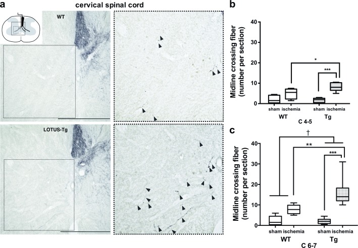
Fig 4. Neuronal remodeling in cortico-spinal fibers.
(a) Representative photomicrographs of the gray matter of the cervical spinal cord in frontal sections of WT mouse and LOTUS-Tg mouse, showing increased midline-crossing fibers (black arrowheads) in LOTUS-Tg mice after MCAO. Insets of the spinal cord scheme indicating the position of the photomicrograph. Images in right panel are shown at higher magnification of indicated dashed boxes. Scale bars indicate 500 μm. (b, c) The number of midline-crossing fibers of WT and LOTUS-Tg mice at the C4-5 and C6-7 levels of the cervical spinal cord. (2-way ANOVA with Tukey post hoc analysis; **p < 0.01, ***p < 0.001, †p < 0.05. Data are interquartile range. n = 9 per group).

