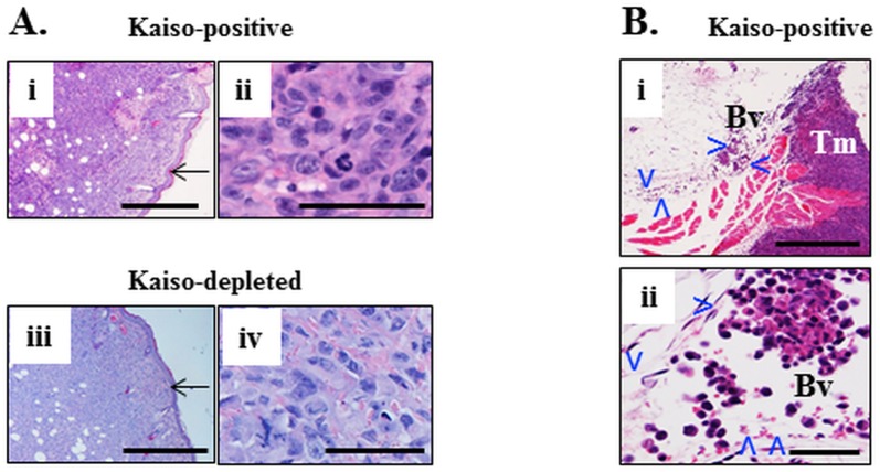
Fig 2. Primary subcutaneous tumors formed by Kaisopositive and kaisodepleted cells with invasion of the lumen of surrounding veins.
Subcutaneous tumor mass of Kaisopositive MDA-231 human mammary carcinoma cells (Ai, ii) and Kaisodepleted tumor cells (Aiii, iv)) implanted into the fat pad of the mammary gland of female NRG mice. Tumor cells abut against the epidermis (arrow in Ai, iii) but do not invade it. Tumor cells are large, markedly pleomorphic, there is high mitotic index. (Bi) A vein (Bv, delineated by arrowheads) is adjacent to the subcutaneous tumor mass (Tm). It is distended by clumps and individual large pleomorphic cells (Bii) and also has scattered red blood cells. H&E. Size bars Ai, ii, Bi– 500 microns; Aii, iv, Bii– 50 microns.

