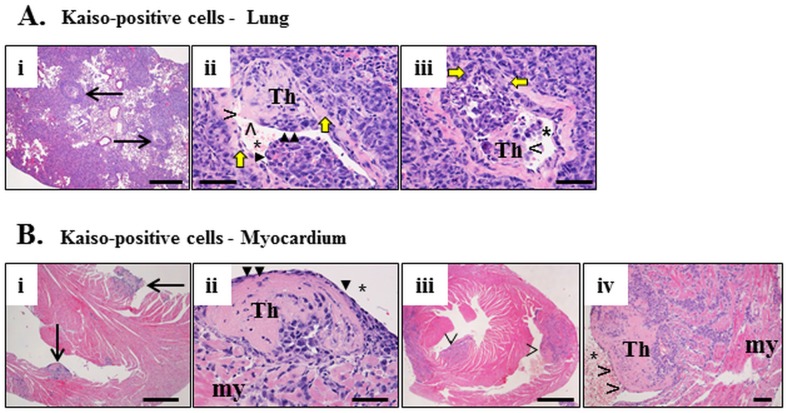
Fig 5. Thrombosis caused by Kaisopositive tumors invading the blood vessels and heart ventricles.
In the lung (A), a number of large blood vessels (two indicated by arrows) have intravascular thrombi delineated from the vascular wall by yellow arrows and protruding in the vascular lumen (Th in Aii, iii). The thrombi are infiltrated by neoplastic cells and are lined by endothelium (solid arrowheads in Aii) or not (open arrowhead in Aiii). In the myocardium (my, B) thrombi protruding into the ventricular lumen (Bi, iii) are also infiltrated by neoplastic cells (Th in Bii, iv) and either lined by endothelium (solid arrowheads in Bii) or not (open arrowheads in Biv). H&E. Size bars; Biii– 1,000 microns, Ai, Bi– 500 microns, Aii, iii, Bii, iv B– 50 microns.

