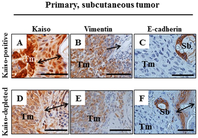
Fig 6. IHC of primary subcutaneous Kaisopositive and Kaisodepleted MDA-231 tumors.
Tumor cells (Tm) of Kaisopositive (A-C) and Kaisodepleted (D-F) masses do not invade the epidermis (double-headed arrow in A, D, F, arrow in B). Kaisopositive tumor cells are labeled strongly positive for Kaiso (A) and vimentin (B) while the Kaisodepleted cells are labelled considerably less (D, E). The labeling with anti-E-cadherin antibody is negative for both types of tumor cells in contrast to the positive labelling of the mouse epithelium in sebaceous glands (Sb in C, F) and in epidermis (F). Size bars A-F– 50 microns.

