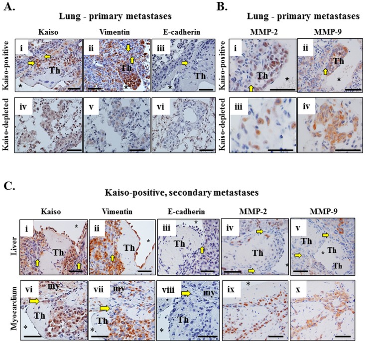
Fig 7. The molecular phenotype of the Kaisopositive MDA-231 cells persist as they metastasize to other distal organs (liver and myocardium).
(A) Neoplastic Kaisopositive cells in lung metastases or thrombi are large, pleomorphic, and stain positive for Kaiso (i) and vimentin (ii), but negative for E-cadherin (iii). In contrast, Kaisodepleted tumor cells are weakly stained for Kaiso (iv) and Vimentin (v) and negative for E-cadherin (vi). (B) Kaisopositive tumor cells in lung metastases also stain strongly for MMP-2 (i), and MMP-9 (ii), while the Kaisodepleted tumor cells stain weakly for MMP-2 (iii), but positive for MMP-9 (iv). The asterisk indicates the lumen of the blood vessel with the thrombus (Th) and the yellow arrows indicate the vascular wall and its obliteration by tumor cells in (A, B). (C) The thrombus (Th) formed in the large blood vessel of the liver (i-v) and in the ventricle of the heart (vi-viii) in mice injected with Kaisopositive cells partially obliterated the vascular wall or endocardium indicated by the yellow arrows (i-viii). Neoplastic cells are stained positive for Kaiso (i, vi), vimentin (ii, vii), MMP-2 (iv, ix), and MMP-9 (v, x), and negative for E-cadherin (iii, viii). Scale bars; A-C– 50 microns.

