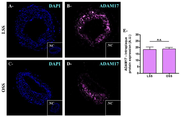
Fig. 2.
ADAM17 expression is not different between LSS- and OSS-induced plaques. A–D) Microphotographs of carotid artery lesion from untreated ApoE−/− mice: LSS-induced plaque showing DAPI (A) and ADAM17 (B); and OSS-induced plaque showing DAPI (C) and ADAM17 (D). The negative control (NC) is shown in the lower right portion of each microphotograph. E) Quantification for ADAM17 expression. n.s.: non-significant (Mann–Whitney nonparametric test). Data were expressed as mean ± SEM.

