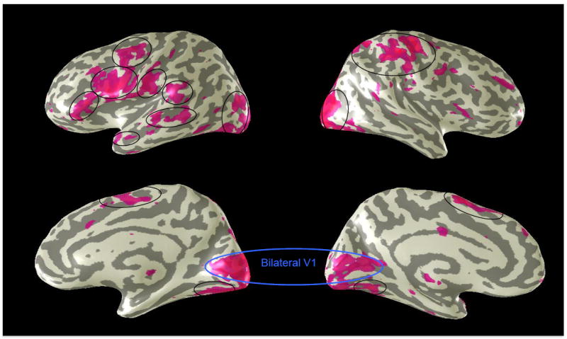
Figure 4. Network for Braille reading.

Cortical regions activated during Braille reading, shown on four views of whole-brain inflated cortex. Note strong lateralization of frontal activations and well-structured activation of V1/2 in the brain of this completely blind individual.
