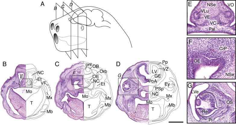
Fig 4. Craniofacial features of newborn S. crassicaudata.
(A) Schematic of a stage 18 dunnart head (postnatal day 0–3) indicating the relative size and position of nostrils, mouth, tongue, eyes and brain, as well as the section planes shown in (B-D). (B-D) Haematoxylin/eosin staining of a rostral-caudal series through the planes depicted in panel A reveals orofacial, sensory and brain structures of newborn dunnarts, including a prominent tongue (T) that encloses the teat inside the mouth (Mo) reaching the lateral margins of the palate (Pal). Large nasal cavities (NC) with fused palate (Pal) and nasal septum (NSe) can be seen, including condensation of mandibular (Mb) and maxillary (Mx) processes, eyes (Ey) fully covered by skin, and scattered vibrissal follicles (VF) in the snout. Telencephalic structures include the olfactory bulb (OB), with surrounding olfactory nerve layer (onl), preplate (Pp), ventricular zone (VZ) and ganglionic eminences (GE) as well as a large lateral ventricle (LV). E-F, inset of the regions highlighted in B-D depicting developing sensory structures. E, the vomeronasal organ (VO) includes a lumen (VLu), neuroepithelium (VE) and capsule (VC) at the base of NSe and Pal. F, a thick olfactory neuroepithelium (OE) is located at the roof of the NC, immediately below the presumptive cribiform plate (CrP). G, eye development has just undergone closure of the lens vesicle (LVe), the pigment epithelium (Pi) outlines a dark ring around cells of the retina (Re). Et, ethmoid bone; Orb, orbital bone; PoA, preoptic area; PSp; presphenoid bone. Scale bar: 500 μm.

