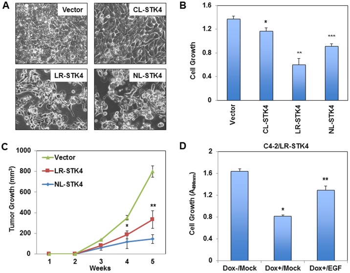
Fig 2. Regulation of C4-2 cell growth by STK4 signaling in all three subcellular locations.
(A) Representative bright field images of C4-2/Vector, C4-2/CL-STK4, C4-2/LR-STK4, and C4-2/NL-STK4 cells. Cell images were captured at 72h post to Dox treatment (4 μg/ml). (B) Growth of C4-2/Vector, C4-2/CL-STK4, C4-2/LR-STK4, and C4-2/NL-STK4 cells in vitro. Cell growth was determined by MTS assay at 72h post Dox exposure. Data (±SD) are the representation of two independent experiments in triplicates, *, **, ***P < 0.007. (C) Prostate tumor xenografts in mice (n = 10 per conditions). C4-2/Vector, C4-2/LR-STK4, and C4-2/NL-STK4 cells were subcutaneously inoculated into the intact nude, immunocompromised male mice. Animals were treated with Dox (0.5 mg/ml) for 6 weeks in drinking water. Tumor sizes were measured weekly for 5 weeks. Tumor growth (volumes) was presented as a function of time, *, **P < 0.01. (D) Growth of C4-2/LR-STK4 cells treated with and without Dox and epidermal growth factor (EGF). Cell growth was determined by MTS assay at 72h post Dox and/or EGF treatment, *, **P < 0.001. Data (±SD) are the representation of two independent experiments in triplicates.

