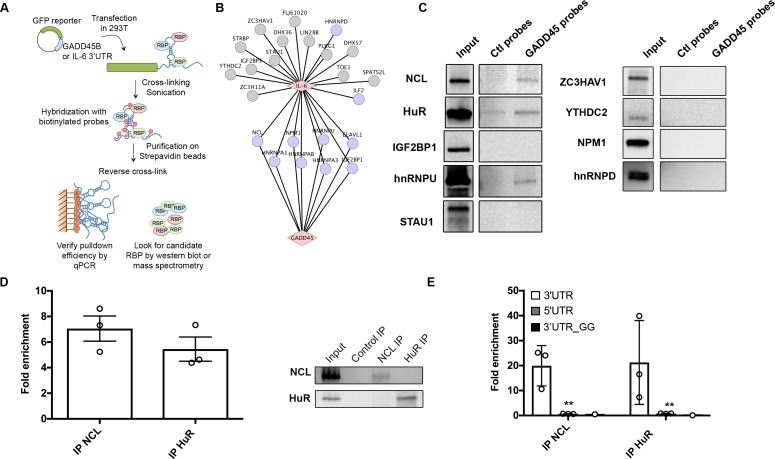
Fig 5. The IL-6 and GADD45B SRE bind a partially overlapping set of cellular proteins.
(A) Diagram outlining the ChIRP assay. (B) Cytoscape network showing the proteins reported to interact with the IL-6 SRE from a previous screen [27] (gray nodes) overlaid with the set of proteins that were also recovered by ChIRP-MS for the IL-6 and GADD45B 3’ UTRs (purple nodes) (C) 293T were transfected with the GFP-GADD45B 3’UTR reporter, then 24h later they were subjected to ChIRP analysis and protein samples were western blotted. (D) Crosslinked lysates of 293T cells were subjected to RNA immunoprecipitation (RIP) with control IgG, anti-NCL, or anti-HuR antibodies and the level of co-purifying endogenous GADD45B mRNA was quantified by RT-qPCR. Bars represent the fold enrichment over the mock IP. (E) 293T cells were transfected with either the GFP-GADD45B 5’UTR, GFP-GADD45B 3’UTR, or GFP-GADD45B 3’UTR_GG reporter for 24 h. Lysates were then subjected to RIP as described in (C). Bars represent the fold enrichment over mock IP.

