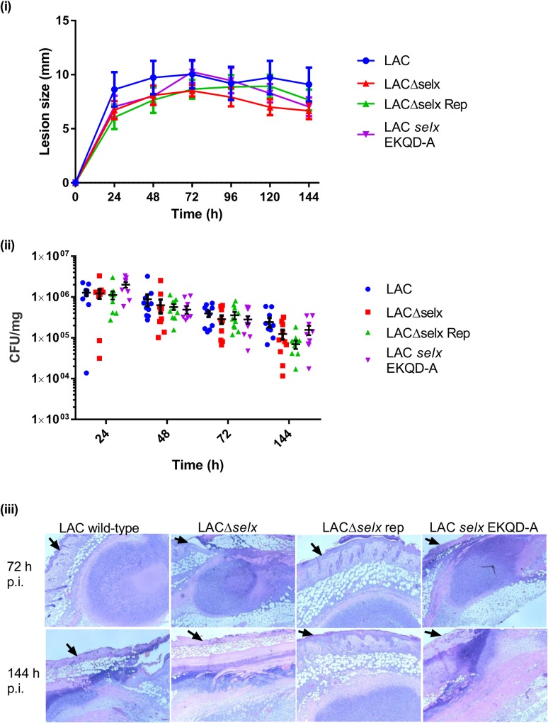
Fig 7. SElX does not contribute to S. aureus virulence in a mouse skin abscess infection model.
(i) Lesion size of each mouse (n = 10) was measured every 24 h post inoculation (p.i.) for 6 d. Mean lesion size ± SEM is plotted for each of the 4 USA300 LAC mutant groups. (ii) Bacteria were recovered from excised skin lesions and enumerated by serial dilutions. CFU were normalised to the weight of tissue homogenised to give the bacterial load per mg of tissue. CFU per mg are displayed for each infected animal; the horizontal line indicates the mean CFU/mg of tissue and vertical bars show the SEM for each group (n = 10) (iii) Representative images from histological examinations of skin lesions 72 h and 144 h post inoculation. Mounted sections were stained with haematoxylin and eosin. Black arrows on each image indicate the surface of the epidermis.

