


Fig. 1. A modular and generalizable design for photoswitchable kinases.
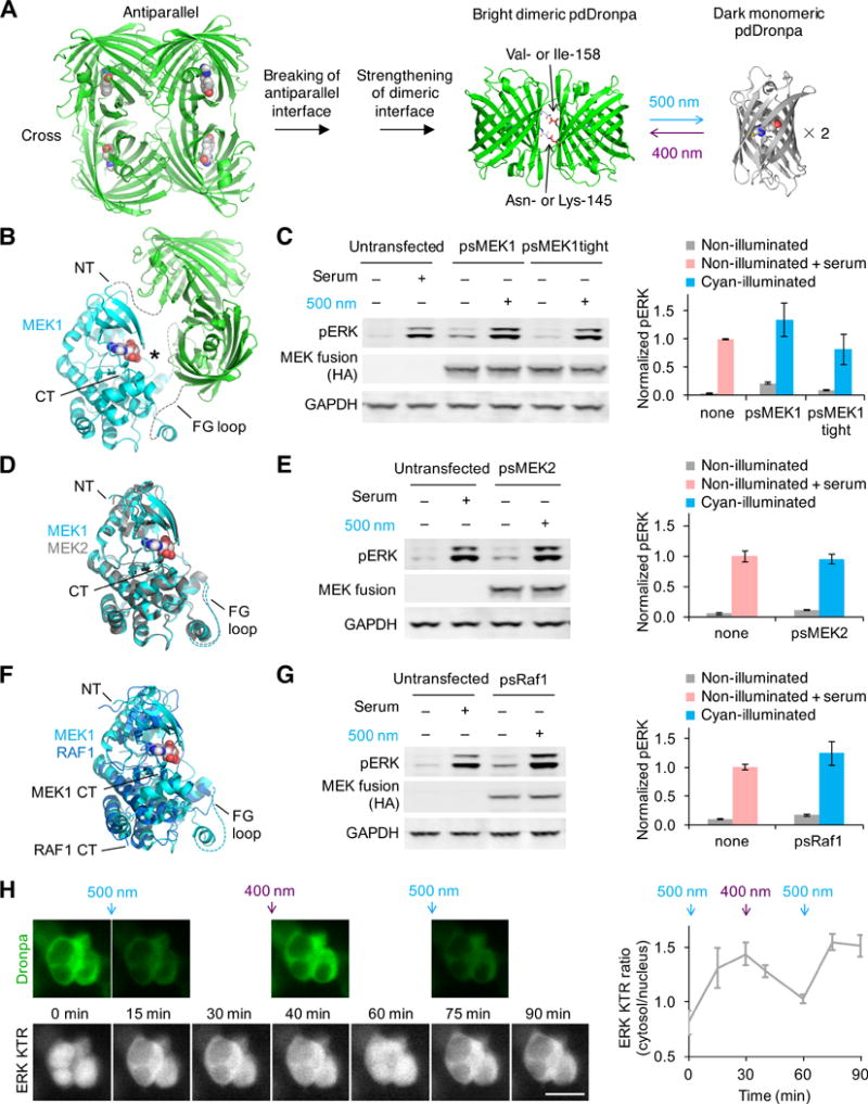
(A) Photodissociable dimeric Dronpa (pdDronpa) variants were engineered from tetrameric DronpaN145. Residues 145 and 158 were further mutated to tune affinity. (B) Structural model of ps(ΔNT)MEK1 in the pre-illuminated state, showing the MEK1 core kinase domain with active site (asterisk) caged by pdDronpa1 domains attached at the NT and the GH loop (rendering based on PDB files 1S9J for MEK1 and 2Z6Y for Dronpa). Note ps(ΔNT)MEK1 contains constitutively activating mutations as well. (C) Light-dependent induction of ERK phosphorylation (pERK) by psMEK1 and psMEK1tight. (D) Structural alignment of MEK1 (PDB 1S9J) with MEK2 (PDB 1S9I). (E) Light-dependent induction of pERK by psMEK2. (F) Structural alignment of MEK1 (PDB 1S9J) with Raf1 (PDB 3MOV). (G) Light-dependent induction of pERK by psRaf1. Note psRaf1 contains a C-terminal CAAX motif for constitutive membrane localization. In (C,E,G), cells were illuminated by 20-mW/cm2 cyan light for 2 min. Protein was detected via an N-terminal HA tag, and lysate loading was monitored by blotting for GAPDH. Serum stimulations were for 5–10 min. Error bars represent standard error of the mean (s.e.m.), n = 3. (H) psMEK1 activation can be temporally and reversibly controlled. Upper panels, intrinsic pdDronpa fluorescence in psMEK1. Lower panels, mRuby2 fluorescence of the ERK KTR sensor. Cells were illuminated with 200-mW/cm2 cyan light for 1 min after the 0- and 60-min timepoints, and with 200-mW/cm2 violet light for 3 s after the 30-min timepoint. pdDronpa fluorescence was imaged immediately after each light stimulation. Scale bar, 20 μm. Chart, quantification of cytosolic/nuclear KTR fluorescence over time. Error bars represent s.e.m. of imaged cells.