
Fig. 4. Optical control of kinase activity in living animals.
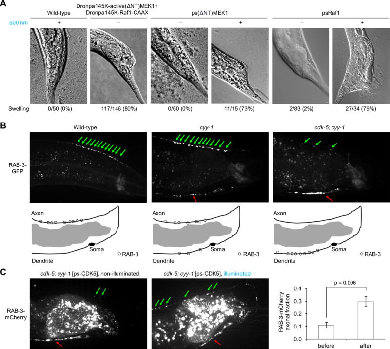
(A) Wild-type C. elegans worms showed no swollen tails even when grown in 500-nm light, while 80% of worms expressing DronpaK145-active(ΔNT)MEK1 and DronpaK145-Raf1-CAAX exhibited swollen tails in the dark (n = 146). 100% of worms expressing psMEK1 in the dark had normal tail shape (n = 50), while 73% of worms expressing psMEK1 in cyan light showed tail swelling (n = 15). Among worms expressing psRaf1, 98% had normal tail shape in the dark (n = 83), while 79% showed tail swelling in light (n = 34). (B) In wild-type worms, synaptic vesicles marked by RAB-3-GFP localize exclusively in the axon. In cyy-1 mutants, RAB-3-GFP is partly mislocalized to the dendrite. In cyy-1;cdk-5 double-mutant worms, nearly all RAB-3-GFP localized to the dendrite. Green and red arrows mark vesicles located in the axon and dendrite, respectively. (C) Light-dependent restoration of CDK5 function in cdk-5;cyy-1 worms. Nearly all RAB-3-mCherry in cyy-1;cdk-5 worms expressing psCDK-5 localized in the dendrite in the absence of light (n = 11), similar to cyy-1;cdk-5 worms. Cyan illumination restored axonal localization of some vesicles (n = 10), similar to the phenotype of cyy-1 worms. The light effect was statistically significant (p = 0.006, unpaired two-sided t-test). Error bars represent standard error of the mean. Worms were exposed to 400-nm light at 0.7-mW/cm2 for 24–48 h before imaging.