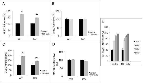
Figure 4. TSP-4 mediates increased adhesion and migration of microvascular EC in response to TGF-β1.
A: TGF-β1-stimulated MLEC from WT and Thbs4−/− (KO) mice were seeded into 24-well plates coated with fibronectin and incubated at 37 °C for 1 h. Unattached cells were removed by washing, and the remaining DNA in the wells was measured using the Cyquant reagent. Adhesion of TGF-β1-stimulated MLEC was compared to the adhesion of non-stimulated cells. *p<0.05 compared to non-stimulated cells, # p<0.05 compared to cells from WT mice; n=3. B: Experiments identical to the ones described in panel A were performed with cultured MAEC; n=3. C: Migration of TGF-β1-stimulated MLEC from WT and Thbs4−/− mice was measured in non-coated Boyden chambers with 8 μm pores and compared to migration of non-stimulated MLEC. DNA in the lower chamber was measured after 4 h at 37 °C. The values are expressed as percent of average migration of non-stimulated cells. *p < 0.05 compared to control (non-stimulated cells), # p<0.05 compared to cells from WT mice; n=3. D: Experiments identical to the ones described in A were performed in MAEC from WT and Thbs4−/− mice. E: Proliferation of TGF-β1-stimulated EC: 12-well plates were coated with fibronectin overnight at 4 °C. EC from WT mice were added and allowed to proliferate at 37 °C for 24, 48, 72, and 96 h; n=3. The amount of DNA in the wells was measured at 24, 48, 72, and 96 h after seeding the cells. The average values of fluorescence are presented as % of average values of fluorescence in control wells with non-stimulated cells.

