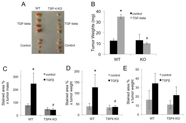
Figure 7. TSP-4 mediates angiogenesis in response to TGF-β1 in a cancer model.
A: Mouse breast cancer EMT6 cells were injected into mammary fat pad of WT or Thbs4−/− (KO) mice. Mice received daily IP injections of TGF-β1. A: representative tumors from one of the experiments. B: tumor weight, n=10, *p < 0.05 compared to control mice injected with PBS; # p<0.05 compared to WT mice. C: Angiogenesis marker CD31 (EC) was visualized by immunohistochemistry in frozen sections of tumors. D: α-actin (SMC) was visualized by immunohistochemistry in frozen sections of tumors from WT and Thbs4−/− mice. E: TSP-4 was visualized by immunohistochemistry in frozen sections of tumors from WT and Thbs4−/− mice. A–D: Mean stained area, % × mean tumor weight, n = 10, *p < 0.05 compared to WT, # p<0.05 compared to WT mice.

