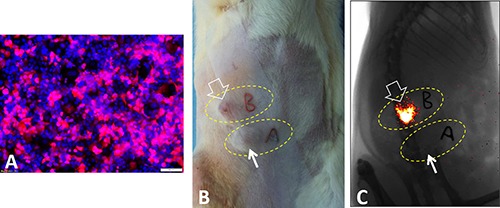
Figure 1.

(A) Fluorescent microscopy shows successful labeling of pancreatic cancer cells by luciferase/m-cherry/lentivirus, which leads to pink-color light emission after administration of luciferin. (B and C) After subcutaneous implantation of the luciferase-labeled pancreatic cancer cells in donor animals, we perform optical imaging to screen bioactive or metabolic active pancreatic cancer tissues, i.e. “bright” tumor tissues that emit luciferase light (open arrows, C), compared to non-bioactive “dark” tumor tissues (short arrows, C). Thus, optical imaging enables us to “sort out” bioactive pancreatic cancer tissues for the next step, orthotopic transplantation in recipient animals.
