Figure 2. title.

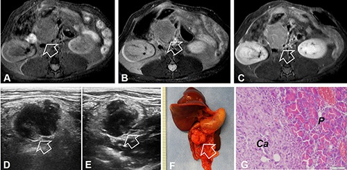
(A–C) MR images of the orthotopic pancreatic head cancer of a recipient rat. The tumor appears as homogeneous hypointense signal on axial T1WI (arrow on A), homogeneous hyperintense signal on T2WI (arrow on B), and heterogeneous enhancement on contrast-enhanced T1WI (arrow on C). (D and E) Ultrasound imaging demonstrates the tumor mass as inhomogeneously hypoechoic intensity on the transvers and longitudinal images (arrows). (F) Photograph of the gross specimen shows a typical pancreatic head tumor (arrow). (G) Histology with H&E staining confirms the formation of the pancreatic ductal carcinoma (Ca) and the normal pancreas (P).
