
Figure 4. Depletion of APOBEC3G attenuated the TGFβ signaling pathway.
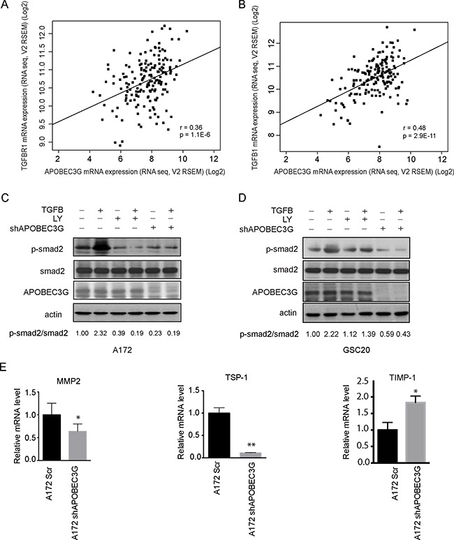
(A–B) TCGA data showed that expression of TGFβR1 (A) and TGFβ1(B) was correlated with APOBEC3G. (C–D) Immunoblotting analysis showed that shRNA targeting APOBEC3G reduced the expression of phosphorylated smad2. A172 cells (C) and GSC20 (D) transfected with scramble or APOBEC3G shRNA, treated with 2.5 ng/ml TGFβ1, 10 mM LY2157299 (TGFβR1 inhibitor), or combination for 24 h, and cell lysis was analyzed by western blot with specific antibodies against phosphorylated smad2, total smad2 and APOBEC3G. The expression of p-smad2 and total smad2 was quantified by Image-J and their ratio was shown in the bottom. (E) The expression of smad2 downstream target genes MMP2, TSP1 and TIMP-1 were analyzed in A172 cells transfected with scramble shRNA (A172SCR) or A712 APOBEC3G knock-down cells (A172 shAPOBEC3G) by quantitative-PCR. Symbols * and ** mean P < 0.05 and p < 0.01, respectively.