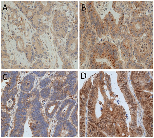
Figure 2. LC3B and p62 immunohistochemical staining in CC tissue.

(A) CC with low LC3B dot like staining. (B) CC with high LC3B dot like staining. (C) CC with both high p62 dot like and high p62 cytoplasmic staining. (D) CC with positive p62 nuclear staining (magnification 20X).
