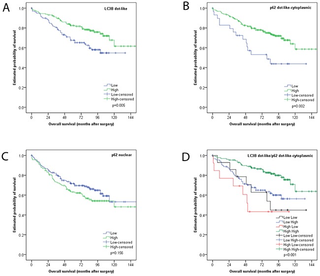
Figure 3. Kaplan Meier survival analysis for LC3B and p62 immunohistochemical staining in CC tissue.

(A) LC3B dot like staining (B) p62 dot like-cytoplasmic staining (C) p62 nuclear staining (D) combination of LC3B dot like/p62 dot like-cytoplasmic staining.
