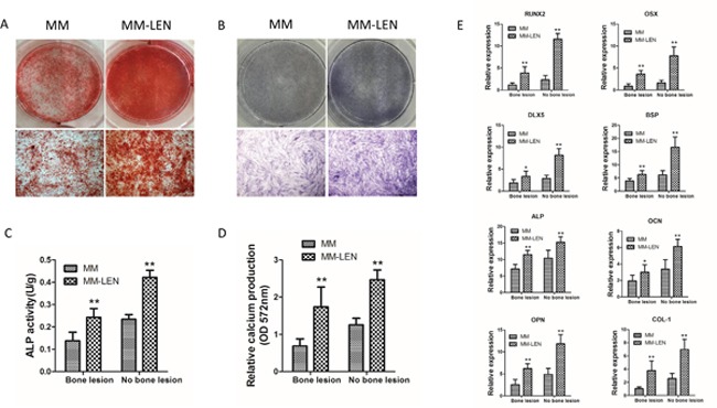
Figure 10. The influence of LEN on impaired osteogenic differentiation of MM-MSCs.

(A) Application of 10 μM LEN in the osteogenic differentiation medium increased mineralization evaluated by alizarin red S staining following 21 days of osteogenic induction. Representative original images and micrographs (original magnification, ×100) of MM-MSCs and MM-MSCs treated with LEN are shown. (B) BMMSCs from MM patients (MM) and MM-patients with LEN treatment (MM-LEN) were evaluated for their osteogenic differentiation potential by ALP staining after 3 days of osteogenic culture. ALP staining was performed with BCIP/NBT. Representative original images and micrographs (original magnification, ×100) of MM-MSCs and MM-MSCs treated with LEN are shown. (C) After 3 days of differentiation in osteogenic medium, ALP activity showed an increase in BMMSCs (derived from MM-B and MM-NB) treated with LEN (n=5, **p< 0.01). (D) Relative calcium production by BMMSCs from MM-B and MM-NB was enhanced with the addition of LEN after differentiation for 21 days (n=6, **p<0.01). (E) Expression of genes, OSX, DLX5, RUNX2, BSP, ALP, OCN, OPN, and COL-1 were upregulated by LEN after osteogenic differentiation for 14 days. The results of MM-MSCs are shown separately as MM-B(n=7) and MM-NB(n=7). All values are expressed as means ± SD. *p < 0.05, **p < 0.01.
