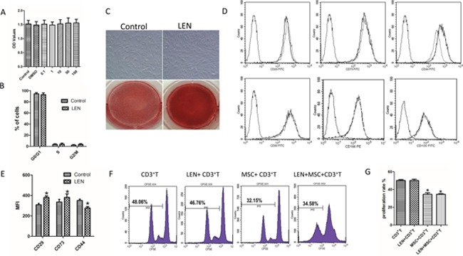
Figure 8. Effects of LEN on proliferation, phenotype, inhibition of T-cell proliferation, and differentiation capacity of BMMSCs.

(A) The proliferative capacity of BMMSCs was measured by CCK-8 assay after being cultured for 3 days in addition with or without LEN. It was not observed significant differences in the distinct HC-BMMSC preparations following LEN treatment (n=11). (B) No significant differences in cell cycle between LEN-treated group and control group were found. (C) LEN treatment did not affect the morphology of MSCs. Calcium production was promoted in the addition of LEN in BMMSCs after differentiation for 21 days. (D, E) LEN addition enhanced the mean fluorescence intensity (MFI) of CD29 and CD73 but suppressed the MFI of CD44 (dotted black line, isotype controls; solid gray line, LEN treated group; solid black line, untreated control;) (n= 12, *p < 0.05). (F, G) LEN addition had no significant effect on the proliferation of CD3+ T cells (p > 0.05). MSCs significantly inhibited the proliferation of CD3+ T cells both without (*p < 0.05) or with LEN addition (*p < 0.05).
