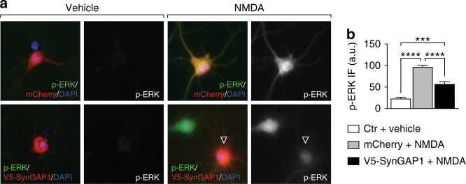
Fig. 8.
Expression of SynGAP1 mitigated NMDA-induced ERK activation in primary neurons. a Primary neurons transiently over-expressing V5-SynGAP1 (red) showed less ERK phosphorylation (p-ERK, green) in response to NMDA exposure, compared to control cells expressing mCherry (red) (or untransfected cells). No p-ERK was detected prior to NMDA treatments. b Quantification of fluorescence intensity of p-ERK signals in transfected neurons showed a significant reduction of ERK activation after NMDA in V5-SynGAP1− compared to mCherry-expressing neurons. Controls (ctr) comprised both mCherry, SynGAP1 or untransfected cells (***p < 0.001; ****p < 0.0001; N = 3; one-way ANOVA (Tukey post hoc)). All error bars are s.e.m

