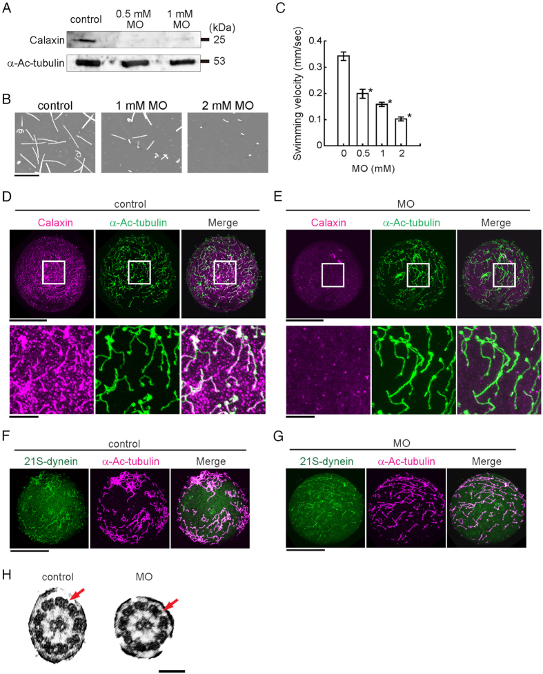
Figure 2.
Knockdown of calaxin results in serious damage of embryonic swimming without changes in ciliary structures. (A) Immunoblots of control or MO-injected embryos (24 hpf) by anti-calaxin and anti-Ac-α-tubulin antibodies. (B) Swimming trajectories of sea urchin embryos at early gastrula stage; images acquired at 0.3 second intervals for 10 seconds are superimposed. Scale, 5 mm. (C) Comparison of mean swimming velocities of embryos injected with different concentrations of calaxin morpholino (MO). N = 61 (control), 38 (0.5 mM MO), 55 (1 mM MO), 28 (2 mM MO). *p < 0.001 vs control (0 mM). (D,E) Immunofluorescence comparison between control (D) and MO (1 mM)-injected (E) embryos. The upper rows, whole embryos; lower rows, enlarged images of squared regions. Scale, 50 μm (upper), 10 μm (lower). (F,G) Control embryo (F) and calaxin morphant (G) immunostained with antibodies against outer arm dynein, showing the presence of outer arm dyneins in morphant cilia. Scale, 50 μm. (H) Thin-section electron microscopy of cilia shows both outer (arrows) and inner arm dyneins in calaxin morphants. Scale, 100 nm.

