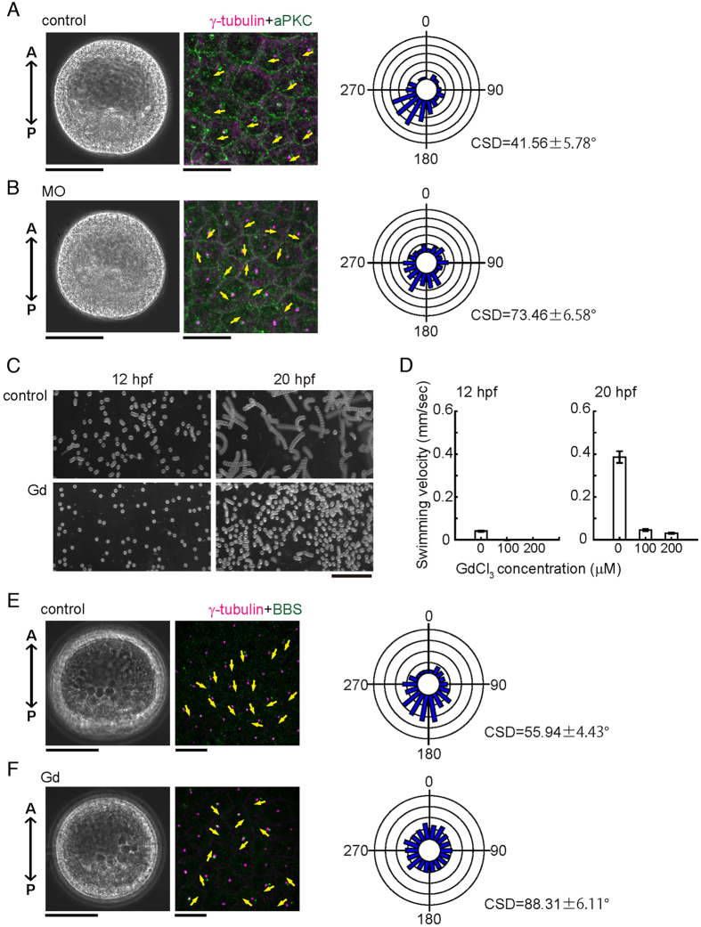
Figure 4.
Calaxin morphants are deficient in the coordinated orientation of ciliary basal structures. (A,B) Phase contrast (left), immunostaining of ciliary basal structures (middle) and circular histograms (right) of control (A) and MO (2 mM)-injected (B) embryos at 24 hpf. Magenta, centrioles (γ-tubulin); green, transition zones (aPKC). Yellow arrows, direction of ciliary basal structures. Circular histograms show the orientation of ciliary basal structures. N = 89 (control) and 95 (2 mM MO), using 7–9 different embryos. Scale, 50 μm (left), 10 μm (middle). (C) Swimming trajectories of embryos treated with 100 μM GdCl3. 10 images acquired at 0.2 second intervals are superimposed. Scale, 1 mm. (D) Comparison of mean swimming velocities of control and Gd3+-treated embryos. N = 30–40 from 3 embryos. (E,F) Phase contrast (left), immunostaining of ciliary basal structures (middle) and circular histograms (right) of control (E) and Gd3+-treated (F) embryos at 20 hpf. Circular histogram, N = 502 (control) and 693 (100 μM Gd3+) using 24–25 different embryos. Scale, 50 μm (left), 10 μm (middle).

