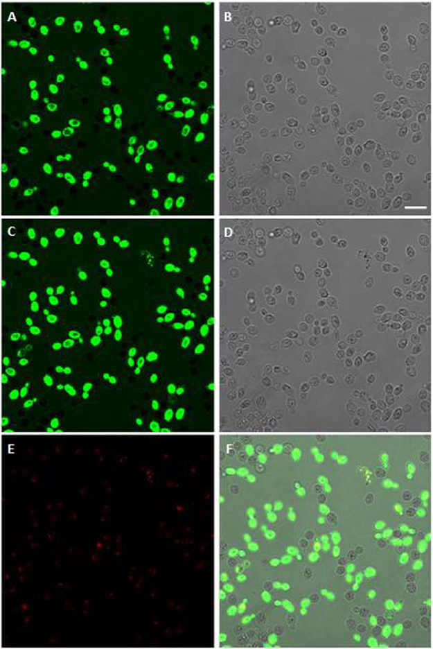
Figure 3.
Internalisation of the L12P peptide into Candida albicans SC5314 cells. Confocal images of living yeast cells incubated in the presence of the fluorescein-labeled peptide for 5 min and 15 min are presented in panels A and C, respectively. The same field is shown by light transmission images in panels B (5 min) and D (15 min). L12P entered into most yeast cells within few minutes; empty vacuoles were seen. After 15 min, some yeast cells were entirely fluorescent and no longer viable as assessed by propidium iodide internalisation (panel E). Panel F: merge of panels C and E. Bar = 10 μm.

