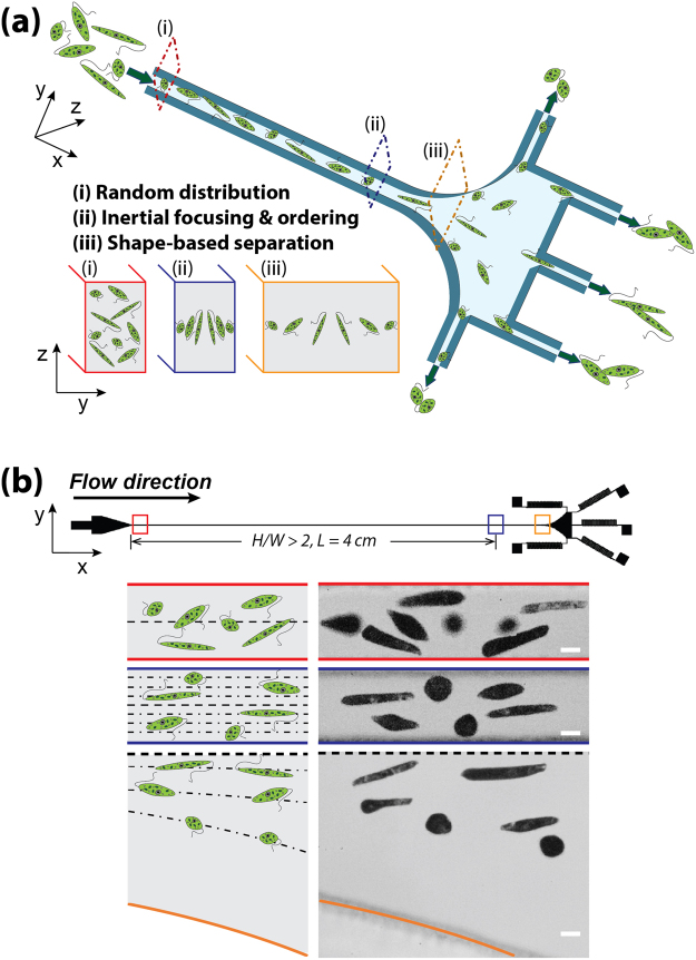
Figure 2.
Shape-based separation of E. gracilis with different aspect ratios due to shape-dependent lateral inertial focusing equilibrium positions. (a) A schematic view of the inertial microfluidic channel and separation principles. (b) A top view (upper) showing the structure and dimensions of the microfluidic device which consists of an inlet, a straight rectangular microchannel, an expansion region, and five outlets with fluidic resistors. Schematics (lower left) and superimposed experimental images (lower right) illustrating the distribution of E. gracilis cells with different ARs at the inlet, downstream, and expansion region. Scale bar = 10 µm.

