Figure 5.
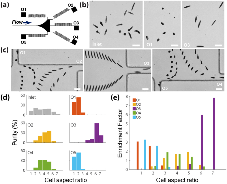
Shape-based separation of E. gracilis cells with different ARs ranging from 1 to 7 at the outlets at Re = 77. (a) An illustration of five outlets with high fluidic resistance (i.e. serpentine channels). (b) Snap-shot pictures comparing E. gracilis proportion at the inlet and outlets. (c) Superimposed experimental images showing that E. gracilis with different ARs are more likely to exit from different outlets: spherical shaped and long rod-shaped cells exit from outlets closer to the sidewall and centerline, respectively. Scale bar = 40 µm (d) A comparison of purity for E. gracilis cells with different ARs ranging from 1 to 7 at inlet and each outlet. (e) Bar graphs of enrichment factors for E. gracilis cells with different ARs for each outlet. At least 100 cells were measured for inlet and each outlet.

