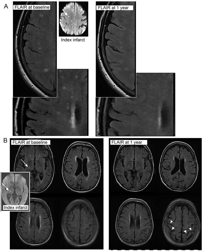
Figure 1. Examples of WMH reduction in 2 participants.
MRIs from patients (A, top and B, bottom) who showed definite visible reduction in WMH on MRIs between presentation with minor stroke (left, baseline; inset, the acute index infarct [arrow] on MRI diffusion tensor imaging) and 1 year (right). Note also the increase in visibility of sulci at 1 year (arrowheads, B, bottom right), indicating a reduction in brain volume accompanying the reduction in WMH volume. FLAIR = fluid-attenuated inversion recovery; WMH = white matter hyperintensities.

