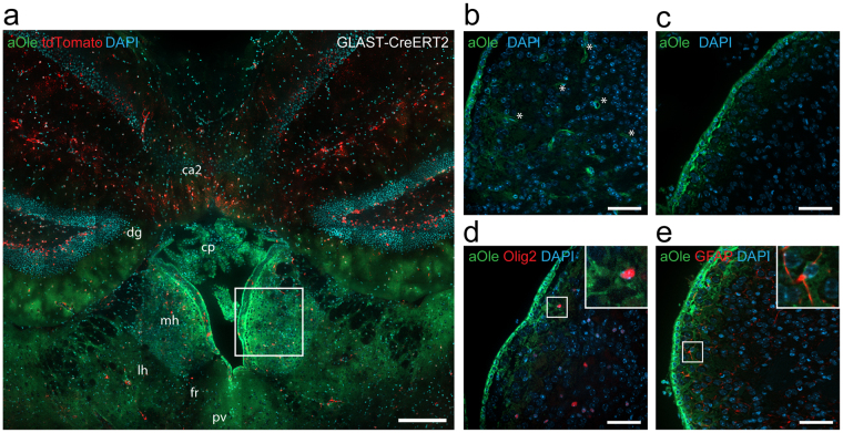
Figure 4.
The alkyne lipid uptake to the medial habenula in vivo corresponds to the in situ observations. (a) In situ cultures of brain slices from of transgenic mice expressing fluorescent tdTomato-reporter after tamoxifen-induced recombination in astrocytes (GLAST-CreERT2) were incubated with 50 µM of alkyne oleate (aOle) for 2 h. Fluorescence microscopy after click-reaction was performed showing alkyne lipids (green), fluorescent protein (red) and nuclei stained by DAPI (blue) in merged channel micrographs. Overview images show medial habenula (mh), lateral habenula (lh), fasciculus retroflexus (fr), paraventricular thalamic nucleus (pv), dentate gyrus (dg), field CA2 of hippocampus (ca2) and choroid plexus (cp) of the dorsal 3rd ventricle. The box indicates the approximated position depicted in panels (b–e). For in vivo uptake into the brain (b) 500 µM of alkyne oleate was continuously applied to the blood circulation of mice for 20 min, or (c–e) 3 mM alkyne oleate was injected as a single dose into the lateral brain ventricle before incubation for 1 h. Fluorescence microscopy after click-reaction was performed and merged channel overview micrographs of the medial habenula are shown. To identify (d) oligodendrocytes and their precursors or (e) astrocytes, samples were immuno-probed for Olig2 or GFAP, respectively. Inserts depict close-up images of individual cells with co-localizing signals. Asterisks indicate a blood vessel. Scale bars, (a) 200 μm; (b–e) 50 μm.

