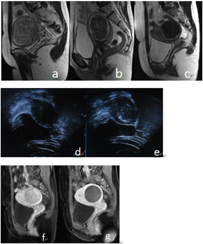
Figure 2.
(a–c) MRI appearance of different types of uterine fibroids on T2-weighted sagittal MRI before treatment; (a) hypointense; (b) isointense; (c) hyperintense. (d–e) US images that show the ablation volume during the real-time USgHIFU; (d) ultrasound image shows a uterine fibroid with hypoecho before treatment; (e) gray scale changes was observed after treatment. (f–g) Enhanced sagittal MRI before and after treatment, the NPV is visible inside uterine fibroids; (f) before treatment, g after treatment.

