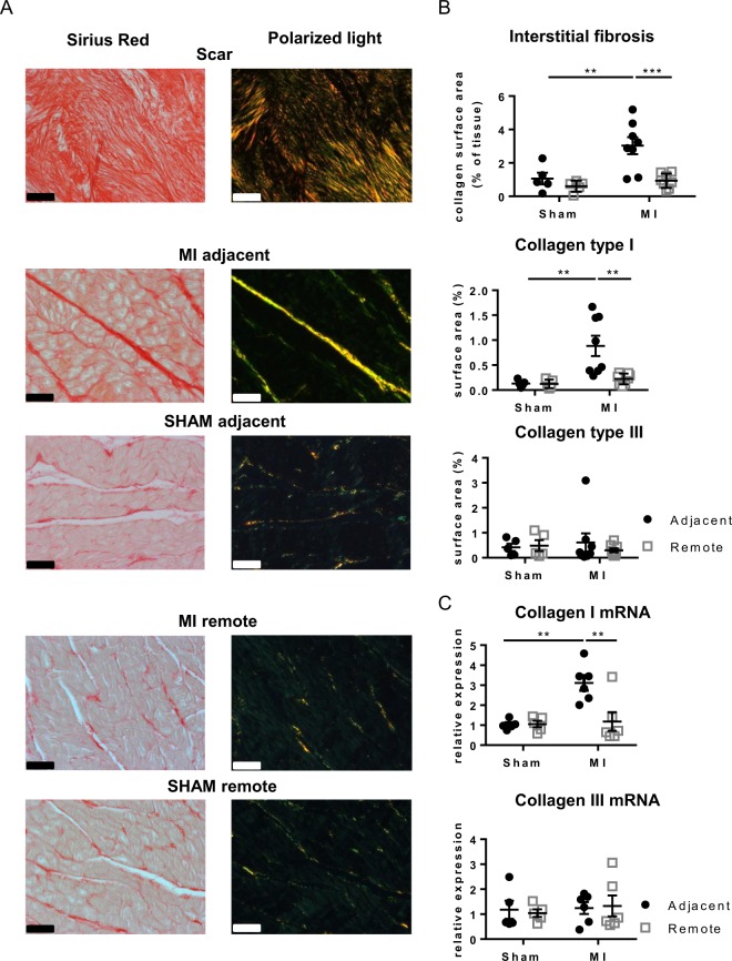
Figure 1.
Interstitial fibrosis in different regions of the LV. (A) Representative images of Sirius red stained sections and polarized light microscopy from the adjacent, remote and scar tissue of SHAM and MI. (B) Analysis of interstitial fibrosis, and in polarized light collagen type I (red-yellow) and collagen type III (green) in SHAM and MI. Scale bars represent 50 µm. (C) Collagen type I and III mRNA expression in the adjacent and remote myocardium of SHAM and MI. (**p < 0.01: ***p < 0.001) (2-way ANOVA with Bonferroni post hoc test).

