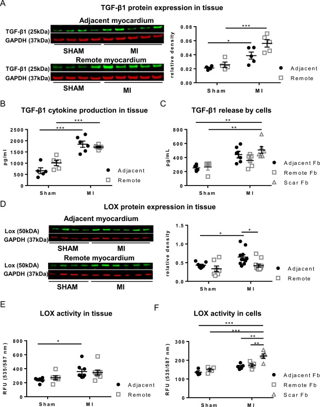
Figure 6.
TGF-β1 and LOX expression during post-MI remodeling. (A) Protein expression by Western blotting of TGF-β1. (B) TGF-β1 concentration measured in cytokine array. (C) TGF-β1 secretion by cultured Fb cells derived from SHAM and MI. (D) LOX expression in tissue from the adjacent and remote myocardium of SHAM and MI. (E) LOX activity in tissue, (F) Lox activity in cultured Fb cells derived from SHAM and MI. *p < 0.05, **p < 0.01. (*p < 0.05: **p < 0.01: *** < 0.001). (2-way ANOVA with Bonferroni post hoc test for 6A, B, D, E;1-way ANOVA with Bonferroni post hoc test for 6 C, F)

