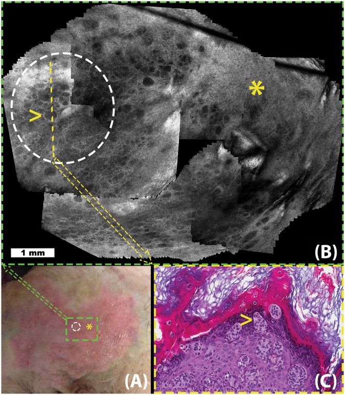
Figure 2.
Extramammary Paget Disease (EMPD): Figure illustrates the use of video-mosaicking for non-invasive examination of tumor spread in a suspected case of EMPD. Panel A (lower left) shows a clinical image exhibiting the suspicious lesion, whose red color and appearance is suggestive of EMPD. In panel B (top), a video-mosaic is obtained by imaging with a handheld confocal microscope in a spiral fashion intended to largely and efficiently cover the green box in panel A. The region inside the white circle on panels A and B was determined to be suspicious in the video-mosaic, and thus a histological section (yellow line in panel B) was excised (shown in panel C). Histological examination of the slice confirmed the findings from the video-mosaic, as illustrated by the high density of tumor cells visible at the yellow arrow in both panels B and C. Unfortunately, during such imaging along a spiral path, the operator could not keep the imaging depth constant and the resulting artifact, as seen in upper left side of the mosaic, occurred at the overlap area. Although the algorithm stitching artifacts through grapcuts based method, such artifacts cannot be completely avoided due to the nature of RCM imaging and challenges in image capture.

