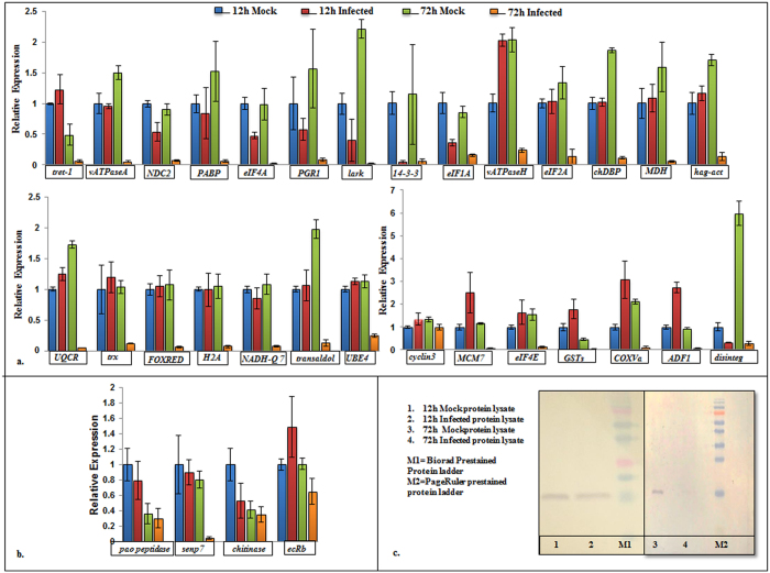
Figure 4.
Comparative expression analysis of proteome data. (a) qPCR based assessment of gene expression in mock-infected and SpltNPV infected Sf21 cells at 12 h and 72 h post infection. The tested Sf21 genes were reportedly down-regulated at 72 h of infection in proteomics analysis. qPCR analysis confirms down-regulation of 27 out of 28 genes. [tret-1: facilitated trehalose transporter, vATPaseA: V-type proton ATPase catalytic subunit A, NDC2: NADH dehydrogenase [ubiquinone] 1 subunit C2, PABP: poly(A)-binding protein, eIF4A: eukaryotic initiation factor 4 A, PGR1: prostaglandin reductase 1, lark: RNA binding protein Lark, 14-3-3: 14-3-3 epsilon protein, eIF1A: eukaryotic translation initiation factor 1 A, vATPaseH: V-type proton ATPase subunit H isoform 2, eIF2A: eukaryotic translation initiation factor 2 A, chDBP: chromodomain helicase DNA binding protein, MDH: malate dehydrogenase, hag-act: heparan-alpha-glucosaminide N-acetyltransferase, UQCR: ubiquinol-cytochrome c reductase, trx: thioredoxin isoform X1, FOXRED: FAD-dependent oxidoreductase domain-containing protein 1, H2A: H2A histone family member V, NADH-Q7: NADH-ubiquinone oxidoreductase Fe-S protein 7, transaldol: transaldolase, UBE4: ubiquitin conjugation factor E4, MCM7: DNA replication licensing factor MCM7, eIF4E: eukaryotic translation initiation factor 4E, GSTs: glutathione S-transferase sigma 1, COXVa: cytochrome c oxidase subunit Va, ADF1: actin-depolymerizing factor 1, disinteg: disintegrin and metalloproteinase domain-containing protein 12]; (b) Relative expression of four Sf21 genes which were found to be up-regulated in proteomics analysis [pao peptidase: pao retrotransposon peptidase family protein, senp7: sentrin/sumo-specific protease senp7, chitinase: chitinase precursor, ecRb: ecdysis triggering hormone receptor isoform B] The genes show similar mRNA levels in both infected and mock-infected samples; (c) Comparative expression of Sf21 protein Histone H3 in mock-infected and infected cells at 12 h (Lane1, 2) and 72 h (Lane 3, 4) post infection through western blot. Blots depicting expression at 12 h (Lane 1,2) and 72 h (Lane 3, 4) were processed separately. The figure shows no change in expression of Histone H3 at 12 h in mock and infected cells but a marked reduction in its expression at 72 h.p.i in SpltNPV infected cells v/s mock-infected cells.

