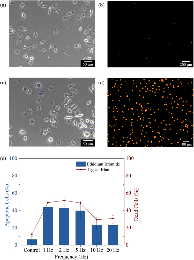
Figure 2.
In vitro cell apoptosis by biaxial pulsed magnetic field. Optical image of HeLa cells and NiFe MNPs (a,b) no field control and (c,d) with magnetic field treatment. The cells (a,c) were stained with Trypan blue (TB) to obtain the dead cells count and (b,d) stained with Ethidium Bromide (EB) to observe apoptotic changes. (e) The quantified data from TB-stained and EB-positive fluorescent cells for frequencies between 1‒20 Hz. The error bars denote the standard deviation across four wells.

