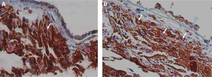
Figure 2.
YAP staining in human choroidal nevi. (A) Nevus 2 with an average of 1.8% YAP-positive cells. A nevus cell with a negative, blue nucleus is seen (white arrow). Magnification of objective × 40. (B) Nevus 12A with an average of 13% YAP-positive cells. Yap+ nuclei in nevus cells are visible (pink, white arrows). A rare RPE cell is found with nuclear YAP staining (blue arrow). Magnification of objective × 40. BM=Bruch’s membrane; cc=choriocapillaris; rpe=retina pigment epithelium.

