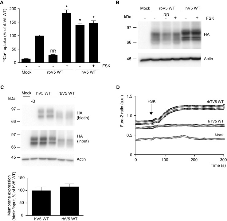
Fig. 1.

Expression and function of human TRPV5. a 45Ca2+ uptake assay of HEK293 cells transfected with either human (hV5 WT) or rabbit (rbV5 WT) wild type TRPV5. Forskolin (FSK, 10 μM) was added directly with 45Ca2+ at the start of the experiment. The uptake is depicted as percentage of rbWT in mean ± SEM (N = 9, from three independent experiments). Asterisk indicates p < 0.05 compared to rbWT. Ruthenium red (RR, 10 μM) is used to define TRPV5-mediated uptake. b Cell lysates of the respective Ca2+ uptake experiments were immunoblotted with HA antibody, using β-actin as loading control. A representative immunoblot is shown. c Cell surface biotinylation of HEK293 cells transfected with human (hV5 WT) or rabbit (rbV5 WT) wild type TRPV5. Samples were analyzed by immunoblotting with HA antibody. The biotin fraction represents the TRPV5 present at the plasma membrane (top panel), and input demonstrates TRPV5 expression in total cell lysates (middle panel), with β-actin as loading control. Representative immunoblot of three independent experiments is depicted. Control without added biotin is indicated as -B. The bottom bar graph depicts the quantified summary of the relative membrane expression compared to input, shown as percentage of hV5 WT. d Averaged Fura-2 ratio in arbitrary units (a.u.) of HEK293 cells expressing mock (n = 51), rabbit TRPV5 wild type (rbV5 WT; n = 69), and human TRPV5 wild type (hV5 WT; n = 101) upon forskolin (FSK) stimulation at t = 60 s indicated by the arrow. The total cell number is obtained in at least three independent experiments
