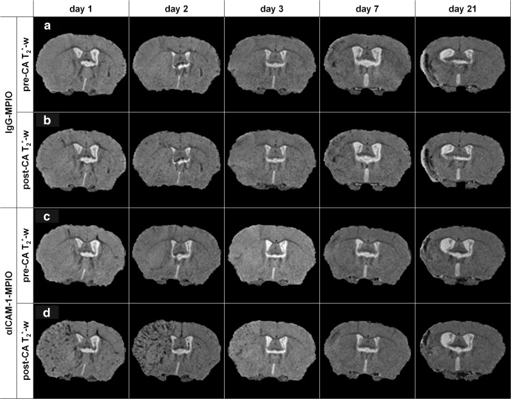
Fig. 3.
In vivo molecular MRI of ICAM-1 expression after stroke. In vivo molecular MRI displayed increased amounts of contrast-enhanced (hypointense) pixels in the lesion territory after injection of αICAM-1-MPIO, but not IgG-MPIO, at days 1, 2, and 3 post-stroke. Ipsilesional hypointense areas on pre-contrast agent (CA) T2*-weighted images were detectable at days 7 and 21, with no further signs of enhancement after contrast agent injection. a, c Pre-CA T2*-weighted (T2*-w) images. b, d Post-CA T2*-w images of a coronal brain slice of animals that received (a, b) IgG-MPIO or (c, d) αICAM-1-MPIO at 1, 2, 3, 7, or 21 days post-stroke

