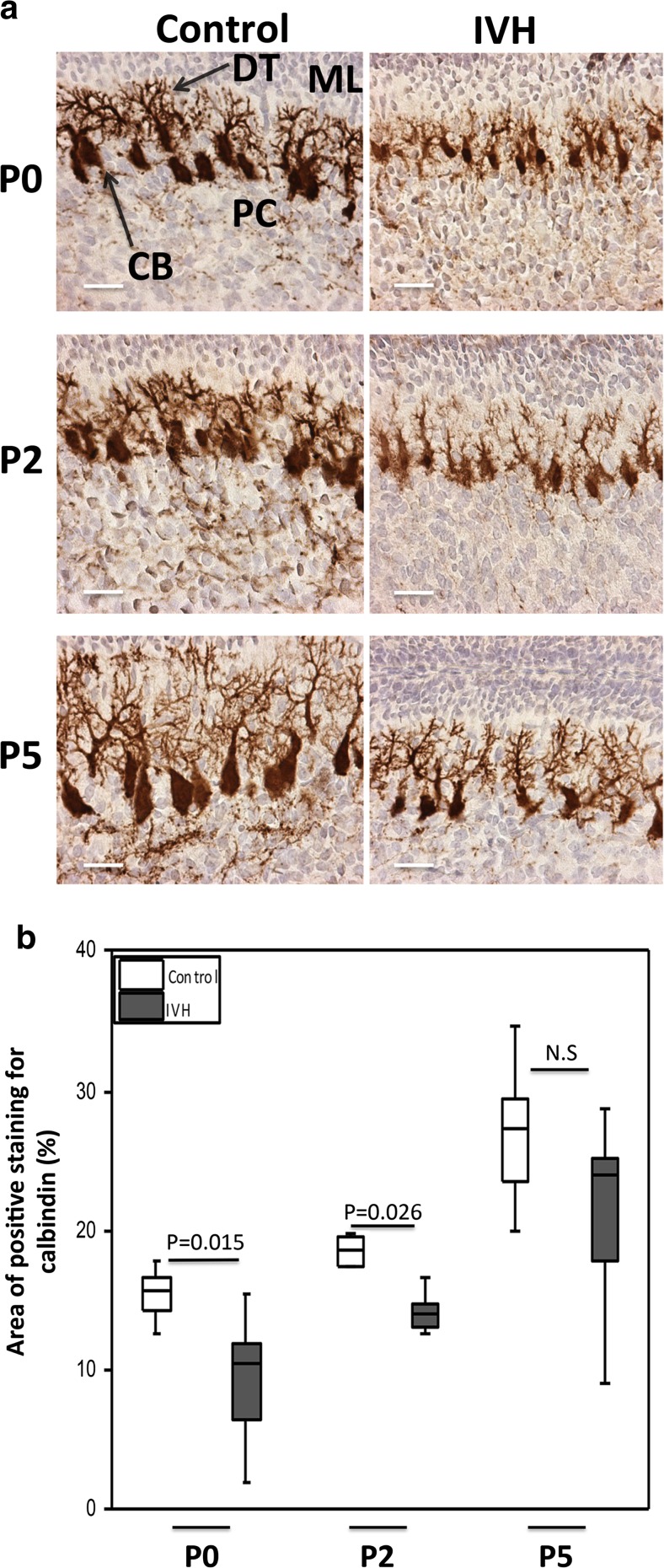
Fig. 4.
Impaired Purkinje cell maturation following preterm IVH. a Immunostaining of calbindin, a calcium-binding protein, was used as a marker of Purkinje cell development in the molecular layer of the developing cerebellum. Calbindin stains are seen as brown to dark brown. Decreased calbindin immunoreactivity was observed in IVH pups (brown) compared to controls (intense dark brown). Observation of neuronal morphology revealed smaller neuronal cell bodies and underdeveloped Purkinje dendrites in IVH pups compared to controls at postnatal time points of P0, P2, and P5. ML molecular layer, PC Purkinje cell, DT dendrites, CB cell bodies; scale bar = 50 μm. b Grading of Purkinje cell development by measurement of percentage area of positive calbindin staining was done in cerebellar tissue sections of both control (white bars; n at P0 = 6, n at P2 = 6, n at P5 = 5) and IVH pups (dark gray bars; n at P0 = 6, n at P2 = 6, n at P5 = 6) at P0, P2, and P5, as described in “Materials and Methods.” Results are presented as box plots displaying medians and 25th and 75th percentiles. Statistical differences between groups for respective time points were analyzed using the Mann–Whitney U test

