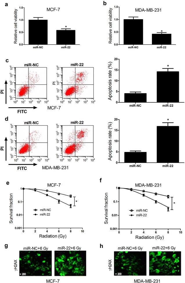
Fig. 2.

Effect of miR-22 overexpression on tumorigenesis and radiosensitivity of breast cancer cells. MCF-7 and MDA-MB-231 cells were transfected with miR-22 or miR-NC and cultured for 48 h. Cell viability in transfected MCF-7 (a) and MDA-MB-231 (b) cells were examined by CCK-8 assay. Apoptosis of transfected MCF-7 (c) and MDA-MB-231 (d) cells was assessed by flow cytometry analysis. Colony formation assay was performed to detect survival fraction in transfected MCF-7 (e) and MDA-MB-231 (f) cells with indicated doses of irradiation (0, 2, 4, 6, or 8 Gy). γ-H2AX foci formation assay was carried out to detect the number of γ-H2AX foci in transfected MCF-7 (g) and MDA-MB-231 (h) cells with 6 Gy radiation. *P < 0.05
