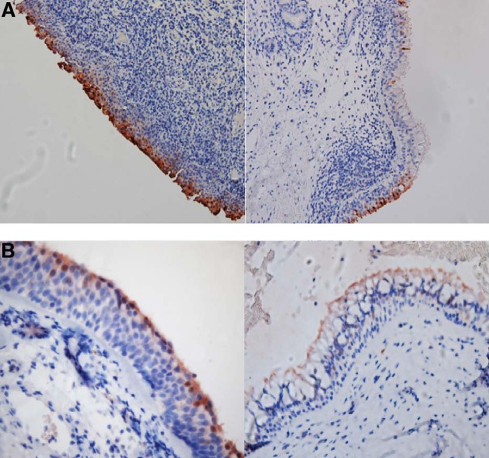
Figure 2.
Immunohistochemical staining for small proline-rich protein 2 (SPRR2) expression in sinus tissue specimens. (A) Low-power view of SPRR2 antibody staining (dark brown) of archived sinus tissue from a subject with chronic rhinosinusitis and/or allergic rhinitis. A representative area of squamous epithelium (left) and ciliated respiratory epithelium (right) demonstrates intense and consistent staining of the squamous epithelium when compared with the respiratory epithelium. (B) High-power view of SPRR2 antibody staining (dark brown) of archived sinus specimens from a healthy control subject with similar findings. A representative area of squamous epithelium (left) and ciliated respiratory epithelium (right) is shown.

