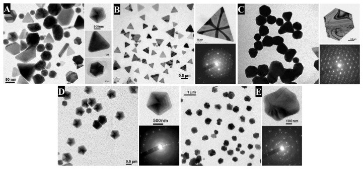
Figure 1.
TEM images of Au NPs synthesized by mixing the cell-free aqueous extract of the fungus Rhizopus oryzae, obtained by tuning the experimental parameters: the amount of HAuCl4, the pH and the reaction time. (A) Mixed nanoplates (triangle, hexagon, pentagon, star, etc.). Right-hand images display individual nanoplates. Scale bars, clockwise from left are 50, 500, 5 and 20 nm. (B) Triangular nanoplates. Scale bars, clockwise from left are 500 and 50 nm. (C) Hexagonal nanoplates. No scale bars were given in the original publication except for 200 nm for the top right figure. (D) Pentagonal nanoplates. All scale bars are 500 nm. (E) Star-shaped nanoplates. Scale bars clockwise from left are 1 µm and 100 nm. For B, C, D and E, the upper picture of each right-hand image corresponds to the high-resolution single-crystalline nanoplates, and the lower image to their corresponding selected area electron diffraction (SAED) patterns, respectively. Reproduced from Reference [36] with permission from Wiley.

