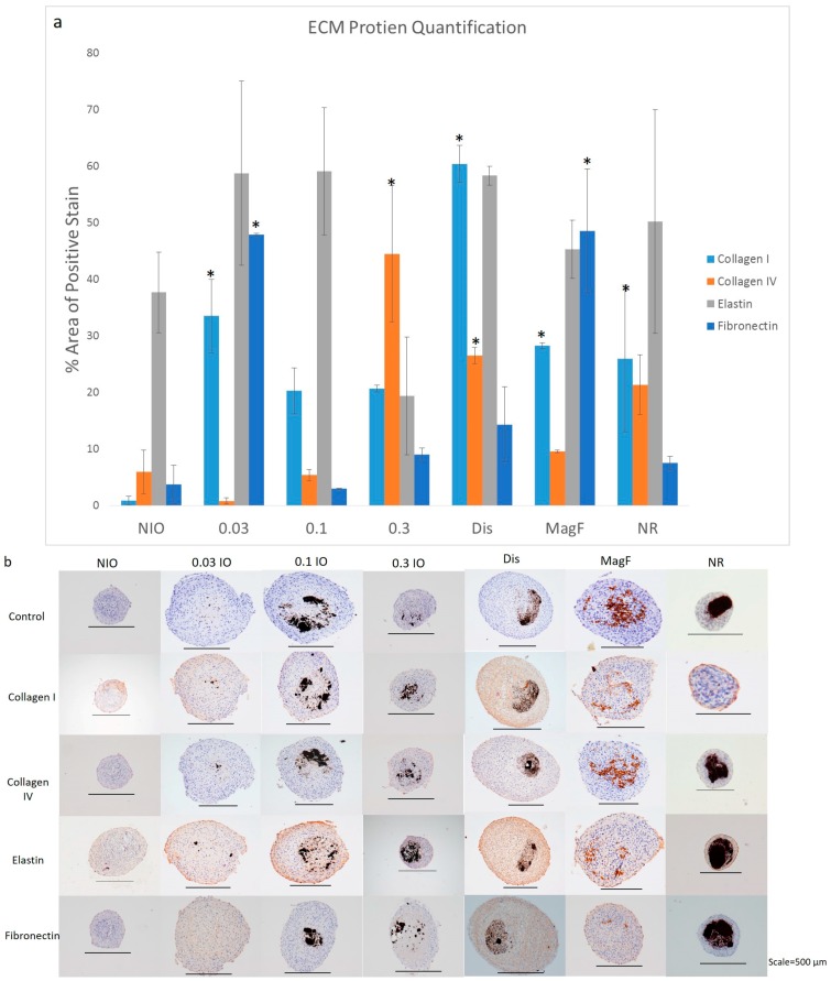
Figure 3.
Immunohistochemistry Staining for MNP spheroids. Spheroids were fabricated with varying MNP concentrations, type, shape, incorporation method, or no MNP. (a) After day 40 time point, spheroids were collected, fixed and processed for immunohistochemical examination for collagen I, collagen IV, elastin and fibronectin. Image J was used to quantify the percent area of positive stain for the various samples. “*” represents samples that have ECM protein production that is significantly different from the NIO control (b) NIO, 0.03 mg/mL IO, 0.1 mg/mL IO, 0.3 mg/mL IO, dispersed (0.3 mg/mL IO), magnetoferritin, and nanorods stained for collagen I, collagen IV, elastin and fibronectin with a control that was not stained.

