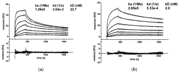
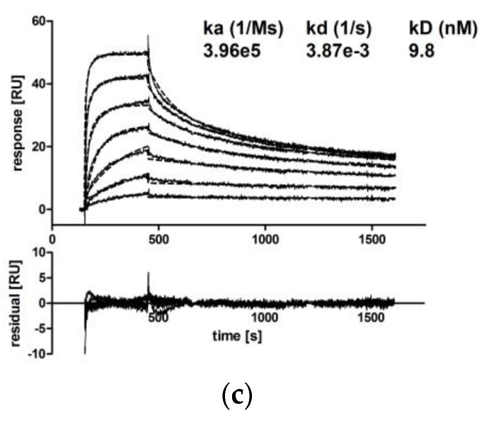
Figure 5.
Kinetic analysis of CM IgA2 monomers, CM IgA2J dimers and CM IgG binding to recombinant EGFR. EGFR was immobilized on one flow cell of a CM5 chip, and a second flow cell served as a reference. Varying concentrations of CM IgA2 monomers (a), CM IgA2J dimers (b) or CM IgG (c) were injected at a flow rate of 50 μL/min for a 5 min association time and followed by a 17 min dissociation time. The difference between the flow cell with immobilized EGFR and the reference flow cell (solid lines) and fits (dotted lines) were plotted. Absolute residuals between data and fits are shown below. One out of two (IgA2 monomers and IgG) independent experiments is shown.


