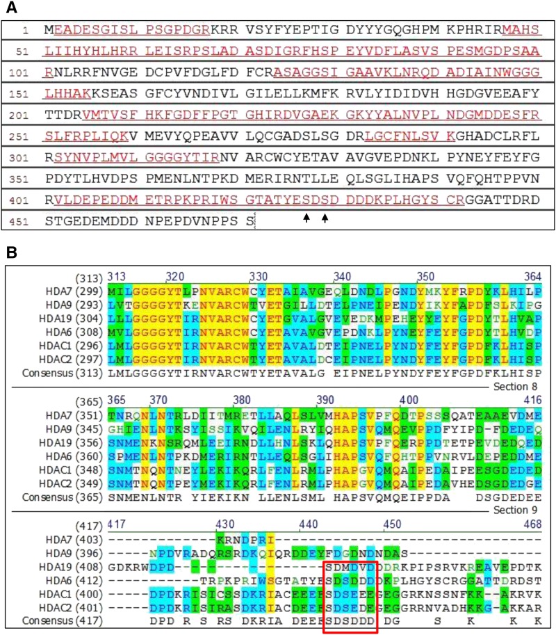
Figure 6.
The Conserved Motif in Phosphorylation Sites among HDA6, HDAC1, and HDAC2.
(A) Peptide sequence of HDA6 detected by LC-MS/MS. The peptides detected by LC-MS/MS are indicated by underlined red letters. The matched peptides exhibit 47% amino acid sequence coverage. Arrows indicate the phosphorylation sites (S427 and S429) reveal by LC-MS/MS.
(B) Amino acid sequence alignment of Arabidopsis HDA6, HDA7, HDA9, and HDA19 with human HDAC1 and HDAC2. HDA6, HDAC1, and HDAC2 contain the conserved motif SDS(E/D)(E/D)(E/D) (boxed in red). Conservative, identical, and blocks of similar amino acid residues are shaded in blue, yellow, and green, respectively.

