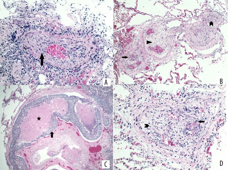
Figure 2.
(A) Pathology of the trans-bronchial biopsy shows arteriole obliteration by smooth muscle proliferation (arrow), consistent with pulmonary vasculopathy. (B) Postmortem lung pathology shows arteriolar smooth muscle proliferation (arrowhead) and fibrosis (triangle) with recanalization of thromboemboli (arrow). Recanalization features multiple small vascular channels containing red blood cells embedded in the fibrotic tissue. (C) Representative pathology of the postmortem lung outlines a distended lymphatic channel filled by tumor thromboembolus (arrow, clumps of blue cells) with necrotic center (star, pink area). (D) Pathology of postmortem lung shows arteriolar myofibroblastic proliferation (arrowhead) in small pulmonary vessels. Also noted are tumor cells (arrow, blue cells with enlarged nuclei) forming thrombi in recanalized lymphatic channels.

