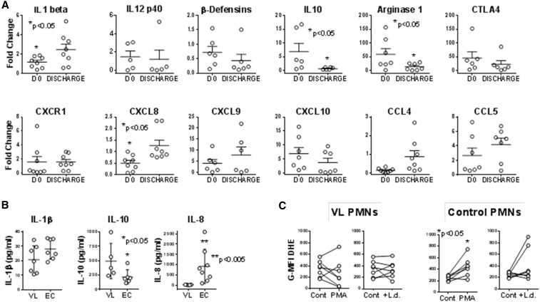
Figure 2.
Neutrophil transcripts, protein, and oxidation. (A) Transcripts. Neutrophils were separated from blood of subjects with visceral leishmaniasis (VL) either prior to VL or after treatment (Tx) or from endemic control (EC) subjects using the Miltenyi CD66abce microbead kit. cDNA was generated and used to quantify transcripts using SYBR green with the primers listed in Table 1. Data show fold change in polymorphonuclear leukocyte (neutrophils) (PMNs) from VL subjects pre- or posttreatment relative to expression of the same gene in PMNs from controls (EC). All CT values were normalized to the abundance of β2 microglobulin as an endogenous constitutively expressed reference. Statistical analyses were done by paired t test. N = 7 VL and seven EC subjects. (B) Proteins secreted by neutrophils were detected by enzyme-linked immunosorbent assay (ELISA). Neutrophils from subjects with VL or ECs were incubated in culture for 6 hours, supernatants were collected and analyzed by ELISA. The data show the amounts of interleukin (IL)-1β, IL-10, or IL-8 protein in supernatants. (C) Oxidation of dihydroethidium (DHE) was documented in neutrophils exposed to stimuli. Isolated PMNs from subjects with acute VL prior to treatment, or from EC subjects were incubated with buffer (Cont), 5 μg/mL phorbol myristate acetate (PMA), or 5:1 opsonized Leishmania donovani promastigotes (+L.d.) for 15 minutes, after which 5 μM DHE (Molecular Probes) was added. DHE oxidation was quantified in CD66b + PMNs by flow cytometry, and plotted as the geometric mean fluorescence index of oxidized DHE fluorescence. N = 7 VL and 7 EC subjects.

