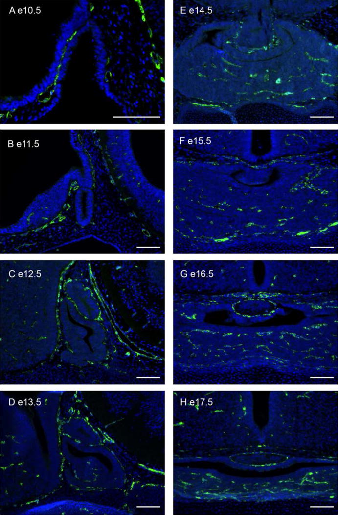
Figure 3. Vascularization of the Developing Pituitary Gland.

(A–H) Immunostaining for CD-31 (PECAM, green) and counterstained with DAPI (blue). Endothelial cells surround the infundibulum at E11.5 and invade the anterior lobe beginning at E13.5. Scale bars 100 μm.
