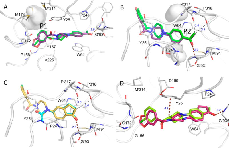
Figure 2.

(A) Comparison of 6 (purple, PDB ID: 4XJP) and 12 (green, PDB ID: 4W1X) in the binding pocket with residues that make substantial contact with the ligands. H-bonds are shown with brown dashed lines. Weaker nonbonded close contacts are illustrated with thin dashed lines. (A) The P1 subsite: the gray surface surrounds a cavity between Met165, Met 174, and Met′314 left unoccupied by ligand or protein atoms following ligand binding. (B) The P2 subsite: unoccupied cavities are also found both above and below the acetylphenyl ring in complexes with 6 and 12. (C) Position of inden-1-one-containing fragment38 (cyan, PDB ID: 4WYF) compared to that of compound 26 (yellow, PDB ID: 4XJO) in the same orientation as panel B. (D) Crystal structures with compounds 36 (red, PDB ID: 5KGS) and 39 (lime, PDB ID: 4KGT).
